Anatomy and physiology of the heart: structure, functions, hemodynamics, cardiac cycle, morphology
The structure of the heart of any organism has many characteristic nuances. In the process of phylogenesis, that is, the evolution of living organisms to more complex ones, the heart of birds, animals and humans acquires four chambers instead of two chambers in fish and three chambers in amphibians. Such a complex structure is best suited for the separation of arterial and venous blood flows. In addition, the anatomy of the human heart implies many tiny details, each of which performs its strictly defined functions.
Heart as an organ
So, the heart is nothing more than a hollow organ, consisting of specific muscle tissue, which performs the motor function. The heart is located in the chest behind the sternum, more on the left, and its longitudinal axis is directed anteriorly, to the left and down. In front, the heart borders on the lungs, almost completely covered by them, leaving only a small part directly adjacent to the chest from the inside. The boundaries of this part are otherwise called absolute cardiac dullness, and they can be determined by tapping the chest wall ().
In people with a normal constitution, the heart has a semi-horizontal position in the chest cavity, in people with an asthenic constitution (thin and tall) it is almost vertical, and in hypersthenics (dense, stocky, with large muscle mass) it is almost horizontal.

heart position
The back wall of the heart is adjacent to the esophagus and to large main vessels (to the thoracic aorta, to the inferior vena cava). The lower part of the heart is located on the diaphragm.

external structure of the heart
Age features
The human heart begins to form in the third week of the intrauterine period and continues throughout the entire period of gestation, passing through stages from a single-chamber cavity to a four-chamber heart.

development of the heart in utero
The formation of four chambers (two atria and two ventricles) occurs already in the first two months of pregnancy. The smallest structures are fully formed by childbirth. It is in the first two months that the heart of the embryo is most vulnerable to the negative influence of certain factors on the expectant mother.
The heart of the fetus is involved in the blood flow through his body, but differs in the circles of blood circulation - the fetus does not yet have its own breathing with the lungs, but it “breathes” through the placental blood. There are some openings in the fetal heart that allow the pulmonary blood flow to be "switched off" from circulation prior to birth. During childbirth, accompanied by the first cry of the newborn, and, therefore, at the time of increased intrathoracic pressure and pressure in the heart of the child, these openings are closed. But this does not always happen, and they may remain in a child, for example, (not to be confused with such a defect as an atrial septal defect). An open window is not a heart defect, and subsequently, as the child grows, it overgrows.
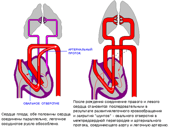
hemodynamics in the heart before and after birth
The heart of a newborn child has a rounded shape, and its dimensions are 3-4 cm in length and 3-3.5 cm in width. In the first year of a child's life, the heart increases significantly in size, and more in length than in width. The mass of the heart of a newborn child is about 25-30 grams.
As the baby grows and develops, the heart also grows, sometimes significantly outpacing the development of the body itself according to age. By the age of 15, the mass of the heart increases by almost ten times, and its volume increases by more than five times. The heart grows most intensively up to five years, and then during puberty.
In an adult, the heart is about 11-14 cm long and 8-10 cm wide. Many rightly believe that the size of the heart of each person corresponds to the size of his clenched fist. The mass of the heart in women is about 200 grams, and in men - about 300-350 grams.
After 25 years, changes begin in the connective tissue of the heart, which forms the heart valves. Their elasticity is no longer the same as in childhood and adolescence, and the edges may become uneven. As a person grows up, and then aging, changes occur in all structures of the heart, as well as in the vessels that feed it (in the coronary arteries). These changes can lead to the development of numerous cardiac diseases.
Anatomical and functional features of the heart
Anatomically, the heart is an organ divided by partitions and valves into four chambers. The “upper” two are called the atria (atrium), and the “lower” two are called the ventricles (ventriculum). Between the right and left atria is the interatrial septum, and between the ventricles is the interventricular septum. Normally, these partitions do not have holes in them. If there are holes, this leads to mixing of arterial and venous blood, and, accordingly, to hypoxia of many organs and tissues. Such holes are called septal defects and refer to.

basic structure of the chambers of the heart
The boundaries between the upper and lower chambers are atrioventricular openings - the left, covered by the leaflets of the mitral valve, and the right, covered by the leaflets of the tricuspid valve. The integrity of the septa and the proper functioning of the valvular leaflets prevent mixing of blood flows in the heart, and promote a clear unidirectional flow of blood.
The atria and ventricles are different - the atria are smaller than the ventricles and have thinner walls. So, the wall of the atria is about only three millimeters, the wall of the right ventricle is about 0.5 cm, and the left one is about 1.5 cm.
The atria have small protrusions - ears. They have a slight suction function for better pumping of blood into the atrial cavity. The mouth of the vena cava flows into the right atrium near its ear, and the pulmonary veins in the amount of four (rarely five) flow into the left atrium. From the ventricles depart the pulmonary artery (more often called the pulmonary trunk) on the right and the aortic bulb on the left.

structure of the heart and its vessels
From the inside, the upper and lower chambers of the heart also differ and have their own characteristics. The surface of the atria is smoother than that of the ventricles. From the valve ring between the atrium and the ventricle, thin connective tissue valves originate - bicuspid (mitral) on the left and tricuspid (tricuspid) on the right. The other edge of the leaflet faces the inside of the ventricles. But in order for them not to hang freely, they are, as it were, supported by thin tendon threads called chords. They are like springs, stretch when the valve flaps close and contract when the flaps open. Chords originate from the papillary muscles from the wall of the ventricles - three in the right and two in the left ventricle. That is why the ventricular cavity has an uneven and bumpy inner surface.
The functions of the atria and ventricles also differ. Due to the fact that the atria need to push blood into the ventricles, and not into larger and longer vessels, they have less resistance to muscle tissue to overcome, so the atria are smaller in size and their walls are thinner than those of the ventricles. The ventricles push blood into the aorta (left) and into the pulmonary artery (right). Conventionally, the heart is divided into right and left halves. The right half serves for the flow of exclusively venous blood, and the left half for arterial blood. Schematically, "right heart" is indicated in blue, and "left heart" in red. Normally, these streams never mix.

hemodynamics in the heart
One cardiac cycle lasts about 1 second and is carried out as follows. At the moment of filling with blood, the walls of the atria relax - atrial diastole occurs. The valves of the hollow veins and pulmonary veins are open. The tricuspid and mitral valves are closed. Then the atrial walls tighten and push blood into the ventricles, the tricuspid and mitral valves open. At this point, there is systole (contraction) of the atria and diastole (relaxation) of the ventricles. After the ventricles have taken in blood, the tricuspid and mitral valves close, and the aortic and pulmonary valves open. Then the ventricles contract (ventricular systole), and the atria fill with blood again. There comes a general diastole of the heart.

cardiac cycle
The main function of the heart is reduced to pumping, that is, to pushing a certain blood volume into the aorta with such pressure and speed that the blood is delivered to the most distant organs and to the smallest cells of the body. Moreover, arterial blood with a high content of oxygen and nutrients is pushed into the aorta, which enters the left half of the heart from the vessels of the lungs (flows to the heart through the pulmonary veins).
Venous blood, with a low content of oxygen and other substances, is collected from all cells and organs from the vena cava system, and flows into the right half of the heart from the superior and inferior vena cava. Further, venous blood is pushed out of the right ventricle into the pulmonary artery, and then into the pulmonary vessels in order to carry out gas exchange in the alveoli of the lungs and to enrich it with oxygen. In the lungs, arterial blood collects in the pulmonary venules and veins, and again flows into the left half of the heart (into the left atrium). And so regularly the heart pumps blood around the body at a frequency of 60-80 beats per minute. These processes are denoted by the concept "Circulation of blood". There are two of them - small and large:
- small circle includes the flow of venous blood from the right atrium through the tricuspid valve into the right ventricle - then into the pulmonary artery - then into the arteries of the lungs - oxygenation of blood in the pulmonary alveoli - the flow of arterial blood into the smallest veins of the lungs - into the pulmonary veins - into the left atrium.
- big circle includes the flow of arterial blood from the left atrium through the mitral valve to the left ventricle - through the aorta into the arterial bed of all organs - after gas exchange in tissues and organs, the blood becomes venous (with a high content of carbon dioxide instead of oxygen) - further into the venous bed of organs - into the system of hollow veins - in the right atrium.

circles of blood circulation
Video: heart anatomy and cardiac cycle briefly
Morphological features of the heart
If you look at sections of the heart under a microscope, you can see a special type of musculature that is no longer found in any organ. This is a type of striated muscle, but with significant histological differences from ordinary skeletal muscles and from the muscles lining the internal organs. The main function of the heart muscle, or myocardium, is to provide the most important ability of the heart, which forms the basis of the vital activity of the whole organism as a whole. Is it the ability to shrink, or contractility.In order for the fibers of the heart muscle to contract synchronously, electrical signals must be supplied to them, which excite the fibers. This is another capacity of the heart – .
Conductivity and contractility are possible due to the fact that the heart autonomously generates electricity in itself. Function Data (automatism and excitability) are provided with special fibers that are an integral part of the conductive system. The latter is represented by electrically active cells of the sinus node, the atrioventricular node, the His bundle (with two legs - right and left), as well as Purkinje fibers. In the case when a patient's myocardial damage affects these fibers, they develop, otherwise called.

cardiac cycle
Normally, an electrical impulse originates in the cells of the sinus node, which is located in the zone of the right atrial appendage. In a short period of time (about half a millisecond), the impulse propagates through the atrial myocardium, and then enters the cells of the atrioventricular junction. Usually, signals are transmitted to the AV node through three main tracts - the Wenckenbach, Thorel and Bachmann bundles. In the cells of the AV node, the time of impulse transmission is extended to 20-80 milliseconds, and then the impulses enter through the right and left legs (as well as the anterior and posterior branches of the left leg) of the His bundle to the Purkinje fibers, and eventually to the working myocardium. The frequency of impulse transmission along all pathways is equal to the heart rate and is 55-80 impulses per minute.
So, the myocardium, or cardiac muscle, is the middle membrane in the wall of the heart. The inner and outer shells are connective tissue, and are called the endocardium and epicardium. The last layer is part of the pericardial sac, or cardiac “shirt”. Between the inner sheet of the pericardium and the epicardium, a cavity is formed, filled with a very small amount of fluid, to ensure better sliding of the sheets of the pericardium at the moments of heart contractions. Normally, the volume of fluid is up to 50 ml, an excess of this volume may indicate pericarditis.

structure of the heart wall and membrane
Blood supply and innervation of the heart
Despite the fact that the heart is a pump to provide the whole body with oxygen and nutrients, it itself also needs arterial blood. In this regard, the entire wall of the heart has a well-developed arterial network, which is represented by a branching of the coronary (coronary) arteries. The mouths of the right and left coronary arteries depart from the aortic root and are divided into branches penetrating the thickness of the heart wall. If these important arteries become clogged with blood clots and atherosclerotic plaques, the patient will develop and the organ will no longer be able to perform its functions in full.

the location of the coronary arteries that supply blood to the heart muscle (myocardium)
The frequency and strength with which the heart beats is influenced by nerve fibers extending from the most important nerve conductors - the vagus nerve and the sympathetic trunk. The first fibers have the ability to slow down the frequency of the rhythm, the latter - to increase the frequency and strength of the heartbeat, that is, they act like adrenaline.

innervation of the heart
In conclusion, it should be noted that the anatomy of the heart may have some deviations in individual patients, therefore, only a doctor is able to determine the norm or pathology in a person after conducting an examination that can most informatively visualize the cardiovascular system.
Video: lecture on the anatomy of the heart
heart, cor, represents a hollow muscular organ that receives blood from the venous trunks flowing into it and drives the blood into the arterial system. The cavity of the heart is divided into 4 chambers: 2 atria and 2 ventricles. The left atrium and left ventricle together make up the left, or arterial, heart, according to the property of the blood in it; The right atrium and right ventricle make up the right, or venous, heart. The contraction of the walls of the heart chambers is called systole. their relaxation - diastole.
The heart has the shape of a somewhat flattened cone. It distinguishes top, apex, base, basis, anterior upper and lower surfaces and two edges - right and left, separating these surfaces.
Rounded apex of the heart, apex cordis, facing down, forward and to the left, reaching the fifth intercostal space at a distance of 8 - 9 cm to the left of the midline; the apex of the heart is formed entirely by the left ventricle. base, basis cordis, facing up, back and to the right. It is formed by the atria, and in front - by the aorta and pulmonary trunk. In the upper right corner of the quadrangle formed by the atria, there is a place - the entry of the superior vena cava, in the lower - the inferior vena cava; now to the left are the places of entry of the two right pulmonary veins, on the left edge of the base - the two left pulmonary veins. Anterior, or sternocostal, surface of the heart, facies sternocostalis. facing forward, up and to the left and lies behind the body of the sternum and cartilage of the ribs from III to VI. The coronal groove, sulcus coronarius, which runs transversely to the longitudinal axis of the heart and separates the atria from the ventricles, the heart is divided into an upper section formed by the atria and a larger lower section formed by the ventricles. Walking on facies sternocostalis anterior longitudinal furrow, sulcus interventricularis anterior. passes along the border between the ventricles, and most of the anterior surface forms the right ventricle, the smaller - the left.
The lower, or diaphragmatic, surface, facies diaphragmatica, adjacent to the diaphragm, to its tendon center. Passes through it posterior longitudinal sulcus, sulcus interventricularis posterior. which separates the surface of the left ventricle (large) from the surface of the right (smaller). The anterior and posterior interventricular sulci of the heart merge with each other with their lower ends and form on the right edge of the heart, immediately to the right of the apex of the heart, heart tenderloin, incisura apicis cordis. The edges of the heart, right and left, are of unequal configuration: the right one is more acute; the left edge is rounded, more obtuse due to the greater thickness of the wall of the left ventricle.

It is believed that the heart is equal in size the fist of the respective individual. Its average dimensions are: length 12-13 cm, maximum diameter 9-10.5 cm, anteroposterior size 6-7 cm. body weight).
Anatomy of the heart (illustrations, 3D images, photos of cuts)
Images and anatomical references
Human heart, anatomy and physiology

human heart is a muscle pump that has been infecting people's minds for hundreds of years. In 2725 BC e. in Egypt, Imhotep came to the conclusion that the pulse is related to cardiac function. In 400 BC e. Hippocrates wrote about the heart as a strong muscle.
In 1628 William Harvey published an explanation of the circulatory process. Between 1857 and 1882, Marey and Dudgeon, independently of each other, created an apparatus for measuring blood pressure when a person was diagnosed with hypertension.
In recent years, molecular biology has helped uncover even more complex features of this engineering masterpiece - human heart. which confirms the words of the psalmist that you and I are “wonderfully made” (Psalm 139:14).
The term "cardiovascular" describes the heart and blood vessels of the body. The blood vessels are also sometimes referred to as the vascular tree, or bed. In this article, we will look at the structure and functions human heart .
The heart is a hollow muscular organ that is located in the central part of the chest, but most of it is located to the left of the midline.
The human heart It consists of two upper chambers called atria and two lower chambers called ventricles. Structurally and functionally, the heart is divided into right and left parts; the right side pumps blood to the lungs, the left pumps blood throughout the body.
The upper chamber, or atrium, collects blood and pumps it into the ventricle, which then ejects it from human heart into large vessels. To ensure blood flow in one direction, each ventricle has inlet and outlet valves.
Left ventricle.
Blood enters the left ventricle from the left atrium through the mitral valve, which consists of two large leaflets that open when the ventricle is relaxed (diastole).
When the filling of the ventricle is completed and it contracts, the force of contraction “presses” the blood against the lower part of the mitral valve leaflets, causing the valve to close. Thanks to this mechanism, blood flows in one direction - from the ventricle to the aorta.
The outlet valve of the left ventricle is called the aortic valve. It has three leaflets, or cusps, that open during ventricular contraction, allowing blood to enter the systemic circulation.
As the ventricle relaxes and the pressure in it falls below the pressure in the aorta, blood begins to flow back (from the aorta to the ventricle).
This backflow of blood causes the leaflets of the aortic valve to fill from above and thus approach each other (touch each other) and slam shut. The valve closes and no backflow of blood into the left ventricle occurs.
Right ventricle.
The inlet valve is a tricuspid valve which, by definition, consists of three leaflets. It provides one-way blood flow from the right atrium to the right ventricle.
Then the blood is ejected into the pulmonary artery through the pulmonary valve (consists of three valves) and flows to the lungs. The tricuspid and pulmonary valves close and open in the same way as the mitral and aortic valves, respectively.
The leaflets of the mitral and tricuspid valves are attached to the walls of the ventricles by "cords" of tissue and muscles called tendon filaments (chords) and papillary (papillary) muscles.
These structures keep the valves from opening in the opposite direction, which would cause blood to flow in the opposite direction. If these leaflets, threads or muscles are damaged due to disease processes, then the valves do not close completely and may "leak" (valve insufficiency).
There are also diseases that cause the valves to narrow, which in turn causes a reduction in blood flow through the valves.
As a result human heart increased resistance is constantly overcome and it increases in size. However, over time, it depletes its reserves of strength and can no longer pump blood as efficiently, which affects the health of the whole body.
The valves can also be affected by both processes at the same time (narrowing and "leaking"), as a result of which the heart function is weakened and blood circulation is disturbed.
cardiac function.
The function of the heart is to pump blood through the systemic circulation (whole body) and through the small (pulmonary) circulation. The right side of the heart pumps blood to the lungs, where carbon dioxide is removed from it and oxygenated.
The left side of the heart pumps blood to the rest of the organs; thus they are supplied with oxygen and nutrients. Waste also enters the blood, but already in the veins, so that later they are removed from the body by organs such as the lungs, kidneys and liver.
The contraction and relaxation of the heart is a cardiac cycle that can be felt by feeling the pulse of blood flowing through the arteries. This can be done by pressing the arteries against the bone, for example, at the wrist, lower leg, neck.
The pulsation of the arteries is created as a result of the buildup of a pressure wave that flows through the human arteries away from the heart and causes a pulsatile expansion of the arterial walls. If we count this pulsation for 60 s, we will get the pulse rate. In a healthy adult, it is approximately 72 beats per minute (the normal range is from 65 to 90).
Each cardiac cycle consists of two phases: diastole and systole.
Diastole (or relaxation of the heart muscle) During this phase, the heart muscle relaxes in order to take some of the flow into the lumen. heart, human blood. The atria then contract to move blood into the ventricles.
The next phase is called systole, or contraction of the ventricles, during which blood is pumped out of the heart. The atria begin to relax in order to take in more blood to repeat the cycle.
You can not only feel the pulse, but also follow the heart cycle by listening to heart sounds through the chest wall using a stethoscope. These sounds are described as "lab-dub", where the first sound "lab" indicates the closure of the mitral and tricuspid valves, and the second sound "dub" - the aortic and pulmonary valves.
Additional sounds usually indicate some kind of heart valve and/or muscle function abnormality. The most common sounds that indicate valve dysfunction are called murmurs.
These sounds are produced when turbulent blood flow occurs due to structural changes in the valvular apparatus. Normally, blood flow is smooth, linear, and non-turbulent (not swirling).
Electrical activity of the human heart.
In order for the heart to beat in an orderly manner, it is equipped with nerve pacemakers (an accumulation of nerve cells in the atria) and a special conduction system that delivers nerve impulses to the heart muscle.
Different parts of the conduction system, and even parts of the heart itself, are capable of beating at different rates. The conduction system provides sequential, coordinated activation from the atria to the ventricles.
This electrical system ensures that the impulses reach all parts of the heart muscle. The electrical axis of the heart is determined according to the electrocardiogram (ECG).
Many of us took up a pen or pencil not only to turn in our drawing homework at school. Sometimes, for one reason or another, in the life of a teenager or already an adult, an inexplicable craving for drawing appears. How you want to pick up a pencil and simply start creating small masterpieces, even if only for yourself or for a close circle of people without claims to world recognition and fame. It may seem that those who perform simple pencil movements on video or in front of you make little or no effort, but in fact this is not the case. Professionalism in drawing, like in any other craft, comes only with experience. Even in the simplest drawings, you can highlight such subtleties and details that you did not even know about before. Now we will look at one of the simplest drawings - a heart. Remember your school years or those moments when we all drew it to each other. This time we will learn how to draw an ordinary heart, with an arrow or wings. It also recommends subscribing. This way you'll be the first to see new content.
Simple Ways
And so, let's figure out how to draw a heart beautifully with a pencil in stages for beginners. All we need is a piece of paper, a simple pencil, and, of course, due perseverance in this endeavor. To make the heart cute, you need to make it symmetrical, and for this, do a couple of simple steps:
Draw two identical circles on a sheet of paper in the same horizontal plane so that both circles intersect slightly. Let's make a reservation right away that the upper halves of the circles will help create beautiful symmetrical edges of the heart. Accordingly, those parts that make up the main picture can be bold, and those that need to be removed can be weaker. It is advisable to draw circles by hand, it's okay if at first the circle does not come out very round, with the development of practical skills this will be corrected. But if the quality of the circles does not suit you initially, you can resort to auxiliary means.
The next figure in the figure is the cross. The vertical line of the cross must pass through the intersection of the circles; to form it, it is enough to draw a line through two points at which the circles from the first point intersect. It does not make sense to lift the vertical line high along the length, its lower part is more useful for the drawing, so do not be stingy and lower it down. In order to understand how low you should lower it, ask yourself: how beautiful to draw a heart, what height proportions will be optimal for you to make the drawing beautiful. The horizontal line is drawn perpendicular to the vertical midpoint of both circles. 
From the extreme points of intersection of the circles with the horizontal line, lower two smooth symmetrical lines to the bottom point of the heart. You should determine the position of this lowest point yourself, as this
parameter, the heart will turn out to be more elongated or more flattened. 
Draw a thick line on the semicircles of each circle upward from the horizontal line to the first point of intersection. 
At this point, drawing the heart is complete. It remains only to remove the extra lines used in the construction and direct the resulting drawing.

Ideal in symmetry and shape, the heart is already in front of you. Of course, this is not the only way to draw a heart.
An easier option for advanced artists
If the previous version seemed boring and not attractive to you due to the presence of a large number of additional constructions, if you need to complete the drawing much faster and there is no way to build circles, if you feel you have sufficient level and skills, we bring to your attention the second method of how to draw a heart pencil step by step. But let's make a reservation right away, you should be good at drawing symmetrical circles, otherwise the heart will turn out to be asymmetrical.
- Divide the sheet into four parts with two perpendicular lines, in other words, depict the same cross.
- Mark on the vertical line the position of the upper and lower points of the heart, and on the horizontal line the same segment to the left and right of the intersection point.
- Connect the upper point with a smooth semicircular line to the leftmost point on the horizontal axis and the same smooth semicircular line to the right point.
- Lower from the extreme left and right points two smooth symmetrical lines to the bottom edge.
For more experienced artists
The next way to draw a heart is even simpler, it will help to draw a heart in just a couple of steps and with the effect of rotation around the axis. But this method is suitable only for experienced professionals who can easily draw symmetrical semicircular lines by hand without using additional constructions.
- draw the most ordinary oval, the edges of which are elongated in a horizontal plane.
- divide the oval with a line in the middle, if the heart should turn out at an angle, the line should be bent in the right direction. Such a frame will show how to draw a heart in stages and quickly.
- select a point just below the top point of the oval on a vertical line and, starting from this position, draw two lines of the upper part of the heart. These lines can completely fit into the oval, or they can protrude beyond it, it all depends on your wishes and vision of the ideal shape for the drawing.
- repeat the previous paragraph with the bottom of the heart - we lower two symmetrical lines to the bottom point.
- add Cupid's arrow.
As a result, we get this picture: 
Let's add details
The drawing can be supplied with additional effects, such as wings, horns, halos, inscriptions, fire and similar additions, giving additional effects and allowing you to harmoniously include the image in one or another motive of the drawing, depending on your ideas. Today we will look at several options for drawing hearts with wings, as the most romantic version of this image. Wings give hearts a special romanticism and sublime tones. It should be noted that the position of the wings in relation to the heart determines the nature of what the author wants to convey: raised to the top, spread wings show firm intentions, pure feelings, desire for a loved one. On the contrary, the more the wings go down (and possibly connect down), the more it shows the heart's attempt to close itself from some external factors and problems, an attempt to hide something under its care and care.
Wings on the heart will tell a lot
So, let's look at how to draw a heart with wings in stages with a pencil. Such a drawing will require you to first study the topic of how to draw a heart or a ready-made template with a heart image. So, for starters, we start from the fact that the finished drawing is already available. It is clear that the easiest and most uncomplicated way is to draw the wings by hand without any skeletons and additional constructions. This method may seem the most common of the above, but at the same time the most difficult, as it will require the author to have practical skills in drawing symmetrical lines and curvilinear shapes by hand. Several variants of the image of wings from the heart should be distinguished. Wings can be depicted from the sides or from the top. The position of the wings themselves in this case does not matter, it is important from which part they grow, so to speak.
Wings with a feather in one row
If you decide to depict wings growing from the top of the heart, then it is better to depict them as small, decorative, this gives a certain sophistication and sophistication when visually contacting the picture. When depicting wings on the sides of the heart, wings outstretched to the sides are an excellent option. We do the following:


Chic wings with a feather in several rows
If you want to get the effect of a heart with huge wings, a span reminiscent of a flying eagle, then it is better to use not a single-level version of the wings, but a multi-level one. The more rows of feathers will be on the wings, the more spectacular the drawing will seem and the nobler the impulse of the heart itself, as if eagle wings carry it towards the beloved. So, let's figure out step by step how to draw a heart with wings with a pencil, depicting a rich pattern of feathers or other additional effects on the wings.
As in previous cases, we begin the image of the drawing with the most common frame of the future heart. It can be depicted in a variety of ways, but it is better to choose one of the above.
To the frame of the heart itself or its finished drawing, we add the frame of future wings. Here you should not skimp on the place, the span and size of the wings themselves should be truly royal. Do not skimp on the place, it is better to draw a smaller heart. Form the frame immediately with several levels: closer to the heart is the smallest, the farthest is the largest. 


Then start drawing each feather, starting with the smallest at the base and ending with the longest and largest at the edges. In principle, you can start applying layers, gradually depicting each row of feathers separately, overlaying the next one without a frame. Repeat the above steps for the second wing.
Want to learn How to draw a human heart with a pencil step by step, follow a few simple steps.
STEP 1. Okay let's start this lesson on the human heart shall we? First draw some guidelines and shapes so that we have a nice workable wireframe to use. Start with a circle for the heart and then draw the bottom of the heart where the heart muscle is located. The three horizontal lines you see drawn will be the plugs on the pulmonary artery, pulmonary veins, and left atrium. The lump you see drawn will be for the aorta.

STEP 2. Okay, draw the actual shape of the aorta as well as the tubes that come from this part of the heart. You will then draw the shape for the pulmonary artery as you see here.

STEP 3. Now, sketch out the shape of the vena cava that is in the vase looking tube, which is all by itself. Next draw of four pipes. The upper tube enters the pulmonary artery, while the last three are the pulmonary veins, which are on the left side. Next sketch out the outer shape of the heart on the left side, which is also part of the heart muscle, and then draw in the veins that lie on the surface of the heart. Lastly you will draw the tubes for the inferior vena cava which is located directly below the lower left side of the heart.

STEP 4. This is your last drawing step and all you have to do is draw out the remaining actual human heart shape and then draw in the superficial veins. Lastly, draw the tubes for the pulmonary vein and the left atrium. Erase all the guidelines that are visible to clear your human heart drawing.