The human brain is an extremely complex system. Thanks to this organ, people have reached the level of development that is observed now. What does he represent?
evolutionary development
In a modern school biology course, topics are considered from simple to complex. First we are talking about cells, protozoa, bacteria, plants, fungi. Later there is a transition to animals and man. To some extent, this reflects the hypothetical course of evolution. Considering the structure, for example, of worms, it is easy to see that it is much simpler than that of humans or higher animals. But these organisms have something important - a ganglion that performs the functions of the brain.
forebrain
If you ask someone to draw the contents of a human skull, most likely, the hemispheres will be schematically depicted. This is indeed one of the most visible and largest parts. But the forebrain also contains the medulla oblongata. In general, their structure is quite complex. And if we take into account a more detailed division, then we can completely name all parts of the forebrain:
- hippocampus;
- basal ganglia;
- big brain.
Of course, there is an even more detailed division, but, as a rule, it is of interest only to specialists. Well, for those who are just expanding their horizons, it will be much more entertaining to find out what all these departments are doing. So what are the functions of the forebrain? And why are there differences between the thinking of right-handers and left-handers?

Functions
The forebrain includes the most recently developed parts. And this means that it is thanks to them that a person has the qualities that he has. And if the diencephalon is mainly engaged in the regulation of metabolism, primitive reflexes and needs, as well as simple motor activity, then the hemispheres are the very place where conscious thoughts are born, where information is learned and memorized, and something new is created.
The hemispheres are also conditionally divided into several parts-zones: parietal, frontal, posterior and temporal. And here are the cells that are engaged, among other things, in the analysis of information coming from outside: visual, auditory, olfactory, gustatory and tactile centers.

The most interesting thing is that from a functional point of view, the left and right hemispheres are different. Of course, there are cases when, in case of damage to one part of the brain, the other took over its tasks, that is, there is a certain interchangeability, but in the usual case, the situation can be as follows: the left hemisphere is engaged in the analysis of the intonation of the speech of another person, and the right hemisphere - the interpretation of the meaning of what was said. That is why left-handers and right-handers, who have more developed different parts, think a little differently.
Also, the functions of the forebrain include memory, various reactions to external stimuli, planning and building future scenarios and situations. There is also a speech center here. All higher nervous activity takes place here: creativity, reflections, ideas.
It is also quite interesting that the forebrain actively develops not only in the prenatal period, but also in the first few years of life. Each new skill and skill, learned word, any important information - all this forms new neural connections. And this kind of card is unique for each person.
- Thinking abilities do not depend on the mass of the brain, but correlate with such a value as the number of convolutions.
- The speed of signals between neurons reaches 288 kilometers per hour. With age, this figure decreases.
- The brain consumes the largest amount of energy among human organs - about 20%. This is a huge figure, given that its mass in relation to the body is only 2%. Also, for its normal operation, a sufficient amount of fluid in the body is necessary.
- The statement that the brain uses only 10% of its resources is a myth. Not so many centers can really work at the same time, but one way or another they are all involved.
text_fields
text_fields
arrow_upward
The diencephalon together with the brain stem is covered from above and from the sides big hemispheres - terminal brain. The hemispheres consist of subcortical nodes (basal ganglia), and have cavities -. Outside, the hemispheres are covered (with a cloak).
Basal ganglia or basal ganglia
text_fields
text_fields
arrow_upward
Basal ganglia or subcortical nodes, (nuclei basales)- the formations are phylogenetically older than the crust. The basal ganglia got their name due to the fact that they lie, as it were, at the base of the cerebral hemispheres, in their basal part. These include the caudate and lenticular nuclei, combined into the striatum, the fence and the amygdala.
Caudate nucleus
text_fields
text_fields
arrow_upward
Caudate nucleus (nucleus caudatus) elongated in the sagittal plane and strongly curved (Fig. 3.22; 3.32; 3.33). Its front, thickened part - head- is placed in front of the thalamus, in the lateral wall of the anterior horn of the lateral ventricle, from behind it gradually narrows and passes into tail. The caudate nucleus covers the visual tubercle in front, from above and from the sides.
Rice. 3.22.
1 - caudate nucleus;
2 - columns of the arch;
3 - epiphysis;
4 - top and
5 - lower colliculus;
6 - fibers of the middle cerebellar peduncle;
7 - pathway of the superior cerebellar peduncle (dissected);
8 - the core of the tent;
9 - worm;
10 - spherical,
11 - cork and
13 - dentate nucleus;
12 - cortex of the cerebellar hemispheres;
14 - superior cerebellar peduncle;
15 - a triangle of a leash;
16 - pillow of the thalamus;
17 - visual tubercle;
18 - posterior commissure;
19 - the third ventricle;
20 - anterior nucleus of the visual mound
 Rice. 3.32.
 Rice. 3.32. Brain - horizontal section through the lateral ventricles:
1 - corpus callosum;
2 - islet;
3 - bark;
4 - tail of the caudate nucleus;
5 - arch;
6 - posterior horn of the lateral ventricle;
7 - hippocampus;
8 - vascular plexus;
9 - interventricular opening;
10 - transparent partition;
11 - head of the caudate nucleus;
12 - anterior horn of the lateral ventricle
Lentil-shaped kernel
text_fields
text_fields
arrow_upward
Lentil-shaped kernel (nucleus lentiformis) located outside of the visual mound, at the level of the island. The shape of the core is close to a trihedral pyramid, with its base turned outward. The nucleus is clearly divided by layers of white matter into a darker-colored lateral part - shell and the medial pale ball, consisting of two segments: internal and external (Fig. 3.33; 3.34).
 Rice. 3.33.
 Rice. 3.33. Horizontal section of the cerebral hemispheres at the level of the basal ganglia:
1 - corpus callosum;
2 - vault;
3 - anterior horn of the lateral ventricle;
4 - head of the caudate nucleus;
5 - inner capsule;
6 - shell;
7 - pale ball;
8 - outer capsule;
9 - fence;
10 - thalamus;
11 - epiphysis;
12 - tail of the caudate nucleus;
13 - choroid plexus of the lateral ventricle;
14 - posterior horn of the lateral ventricle;
15 - cerebellar vermis;
16 - quadrigemina;
17 - posterior commissure;
18 - cavity of the third ventricle;
19 - pit of the lateral furrow;
20 - islet;
21 - anterior commissure
 Rice. 3.34.
 Rice. 3.34. Frontal section through the cerebral hemispheres at the level of the basal ganglia:
1 - corpus callosum;
2 - lateral ventricle;
3 - caudate nucleus (head);
4 - internal capsule;
5 - lenticular shaped nucleus;
6 - lateral furrow;
7 - temporal lobe;
8 - fence;
9 - islet;
10 - outer capsule;
11 - transparent partition;
12 - radiance of the corpus callosum;
13 - cerebral cortex
Shell
text_fields
text_fields
arrow_upward
 Rice. 3.35.
 Shell (putamen) genetically, structurally and functionally close to the caudate nucleus.
Both of these formations have a more complex structure than the pale ball. They are approached by fibers mainly from the cerebral cortex and thalamus (Fig. 3.35).
Rice. 3.35. Afferent and efferent connections of the basal ganglia:
1 - precentral gyrus;
2 - shell;
3 - outer and inner segments of the pale ball;
4 - lenticular loop;
5 - reticular formation;
6 - reticulospinal tract,
7 - rubrospinal tract;
8 - cerebellar-thalamic tract (from the dentate nucleus of the cerebellum);
9 - red core;
10 - black substance;
11 - subthalamic nucleus;
12 - Zona incerta;
13 - hypothalamus;
14 - ventrolateral,
15 - intralaminar and centromedian nuclei of the thalamus;
16 - III ventricle;
17 - caudate nucleus
pale ball
text_fields
text_fields
arrow_upward
The globus pallidus (globus pallidus) is mainly associated with the conduction of impulses along numerous descending pathways to the lower brain structures - the red nucleus, the black substance, etc. The fibers from the neurons of the pallidus go to the same thalamic nuclei that are associated with the cerebellum. From these nuclei, numerous pathways lead to the cerebral cortex.
The pale ball receives impulses from the caudate nucleus and the putamen.
The striatum (corpus striatum) (striatum), which combines the caudate and lenticular nuclei, belongs to the efferent extrapyramidal system. The dendrites of striatal neurons are covered with numerous spines. They terminate the fibers from the neurons of the cortex, thalamus and substantia nigra (Fig. 3.35). In turn, the striatal neurons send axons to the intralaminar, anterior, and lateral nuclei of the thalamus. From them, the fibers go to the cortex, and thus the loop closes. feedback between cortical neurons and striatum.
In the process of phylogenesis, these nuclei were built over the nuclei of the midbrain. Receiving impulses from the thalamus, the striatum takes part in the implementation of such complex automatic movements as walking, climbing, running. In the nuclei of the striatum, the arcs of the most complex unconditional ones are closed, i.e. congenital, reflexes. The extrapyramidal system is phylogenetically older than the pyramidal system. In a newborn, the latter is still not sufficiently developed and impulses are delivered to the muscles from the subcortical ganglia through the extrapyramidal system. As a result of this, the child's movements in the first months of life are characterized by generalization, non-differentiation. As the cerebral cortex develops, the axons of their cells grow to the basal ganglia, and the activity of the latter begins to be regulated by the cortex. The subcortical ganglia are associated not only with motor reactions, but also with vegetative functions - these are the highest subcortical centers autonomic nervous system.
amygdala
text_fields
text_fields
arrow_upward
amygdala (corpus amygdaloideum) (amygdala) - accumulation of cells in the white matter of the temporal lobe. With help anterior commissure it connects with the body of the same name on the other side. The amygdala receives impulses from a variety of afferent systems, including the olfactory system, is related to emotional reactions(Fig. 3.36).
 Rice. 3.36.
 Rice. 3.36. Brain structures associated with the amygdala: afferent (A) and efferent (B) connections of the amygdala:
1 - nuclei of the thalamus;
2 - periaqueductal gray matter;
3 - parabrachial nucleus;
4 - blue spot;
5 - suture cores;
6 - the core of a single path;
7 - dosal nucleus X nerve;
8 - temporal cortex;
9 - olfactory cortex;
10 - olfactory bulb;
11 - frontal cortex;
12 - cingulate gyrus;
13 - corpus callosum;
14 - olfactory nucleus;
15 - anterior-ventral and
16 - dorsomedial nuclei of the thalamus;
17 - central,
18 - cortical and
19 - basolateral nucleus of the amygdala;
20 - hypothalamus;
21 - reticular formation;
22 - partition;
23 - black substance;
24 - ventromedial nucleus of the hypothalamus; XXIII, XXIV, XXVIII - cortical fields
Brain (continued)
The greatest size and complexity in mammals reaches the anterior, or terminal, brain (telencephalon), consisting of two cerebral hemispheres (hemispheri cerebri). Apparently, the hemispheres arose mainly (and perhaps exclusively) in connection with olfactory reception. Smells do not mean so much in the life of higher primates, including humans. However, at earlier stages of evolution, up to the ancestors of vertebrates, the sense of smell was main channel through which animals receive information about the world around them. Therefore, it is quite natural that the olfactory centers of the brain served as the basis on which complex nervous mechanisms subsequently developed. Already on early stages evolution of tetrapods of the hemisphere turn into large and important centers of correlation of sensory signals. By the time when mammals appeared, the strongly expanded surface of the hemispheres became the dominant associative center, the place of localization of higher mental activity. In different representatives of the class, the ratio of the mass of the forebrain hemispheres to the mass of the entire brain varies: in the hedgehog ( Erinaceus europaeus) it is 48%, for proteins ( Sciurus vulgaris) - 53%, wolf ( canis lupus) - 70%, in the common flank ( Delphinus delphis) - 75%, in most primates - 75-80%, in humans - about 85%. In birds, the large hemispheres roughly correspond in mass to the rest of the brain or are inferior to it, sometimes several times. Finally, the extreme importance of the cerebral hemispheres is evidenced by the fact that their destruction leads to the complete functional failure of the mammal.
From below, the olfactory bulbs (bulbi olfactorii) adjoin the anterior part of the hemispheres. These formations are most developed in animals with a good sense of smell and are greatly reduced in purely aquatic forms. According to the difference in their development, several types of brain structure are distinguished. In marsupials, insectivores, edentulous, carnivores, rodents, and some others, the olfactory bulbs are large and protrude well when looking at the brain from above. This type of brain with the perfect development of the olfactory lobes is called macrosmatic. In pinnipeds, sirens, and many primates, the bulbs are poorly developed; These animals have a microsmatic brain. Finally, for cetaceans, the so-called. anosmatic brain with reduced olfactory bulbs. It used to be thought that the ability to distinguish between chemical signals in whales and dolphins was completely lost, but it turned out that this was not entirely true.
The surface layers of the hemispheres of the forebrain of mammals form the pallium, or the cerebral fornix (pallium). The upper layer, consisting of the bodies of neurons and non-fleshy nerve fibers, is called the cortex (cortex cerebri) and is the gray matter of the fornix. The bodies of neurons are located in the cortex in layers, forming a kind of screen structures. This organization of the brain allows you to spatially display the outside world based on information coming from the senses. Screen structures are characteristic of the most important brain centers of mammals, while in other vertebrates they are less common, mainly in the visual centers. Under the cortex is a layer of myelinated processes of neurons - the white matter of the fornix. The nerve fibers of the white matter form conductive bundles, soldering the hemispheres - cerebral commissures. Although the main part of gray matter is concentrated in the cortex, it is also present in the subcortical layer, where it is represented by relatively small clusters - nuclei. These include striatal bodies (corpora striata), which lie under the cavity of the lateral ventricle and got their name because of the nerve fibers crossing them. Under the control of the cortex, the striatum perform the function of regulating stereotyped, automated reactions - unconditioned reflexes.
Schemes reflecting the progressive development of the forebrain hemispheres. Side view of the hemisphere with the olfactory bulb. Different departments that differ from each other cytologically are stained differently.
A - the hemisphere is just the olfactory lobe.
B - the dorsal section is differentiated - the archipallium (= hippocampus) and the ventral section - the basal nucleus (striatum).
B - the basal nuclei moved to the inner region of the hemisphere.
G - a small area appears, which is a neopallium.
E - the paleopallium is pushed back to the medial surface of the hemisphere, but the neopallium is still modest in size, and significantly developed olfactory regions remain under the olfactory groove.
E - the primitive olfactory region is preserved only in the ventral region, and the neopallium reaches an extremely strong development. (According to Romer and Parsons, 1992.) The fornix of the brain began to form even in fish. In connection with the progressive development of the scent sensory system, they have a paleopallium, or an ancient vault (paleopallium), which completely covers the small hemispheres. At the stage of lobe-finned fish, in the dorsal part of the hemispheres, closer to the axis of the body, the archipallium, or old vault (archipallium), appears. In amphibians and primitive reptiles, he gets further development, as a result of which the paleopallium is pushed aside and preserved only along the lateral surface of the hemispheres. At the same time, the paleopallium continues to have a predominantly olfactory character and, at the highest stages of evolution, forms the olfactory lobes of the hemispheric cortex. The archipallium is to a certain, albeit small, degree a correlative center, receiving ascending fibers from the diencephalon, as well as fibers from the olfactory bulb and olfactory lobe; it also appears to be related to emotional behavior. The nerve pathway from this area to the hypothalamus is the main element of the fiber bundle, called the fornix in mammals.
In amphibians, for the first time, the rudiment of a neopallium, or a new vault (neopallium), appears. In reptiles, the neopallium already forms a small area between the ancient and the old vault. From the very beginning of its evolution, this area has been an associative center that, like the basal nuclei, receives fibers that switch sensory signals from the brain stem to it and, conversely, transmit commands directly to the motor columns.
In monotremes, the neopallium is still wedged between the paleopallium on the outside and the archipallium on the inside. In marsupials, it grows beyond the roof and side walls of the hemispheres. At the same time, the archipallium is forced out to the medial surface, and the paleopallium is limited to the ventrolateral part of the hemisphere, located below the nasal groove (fissura rhinalis) - a groove that represents the border between the olfactory and non-olfactory areas of the cortex. In placental, due to the further complication and expansion of the neopallium, the hemispheres reach such a size that they exceed the volume of the rest of the brain combined. The hemispheres grow back and to the sides, successively covering the diencephalon, midbrain and part of the cerebellum. Paired ventricles and ancient structures responsible mainly for the sense of smell (olfactory bulbs, old layers of the fornix and the nerve bundles and nuclei associated with them) are pushed aside and deformed in the placental brain. Thus, the paleopallium is preserved on the ventral surface of the hemisphere in the form of a small olfactory area called the pear-shaped lobe (lobus piriformis), and the archipallium is rolled up in the depth of the fold of the temporal lobe into a roll called the hippocampus. The superiority of the cerebral hemispheres over other parts of the brain is noticeable in all mammals, but it is especially pronounced in advanced forms, such as humans. The hemispheres also dominate functionally. In contrast to mammals, the complication of the forebrain in birds is expressed mainly in the growth of the basal nuclei (nuclei basales), and not the arch remaining thin. 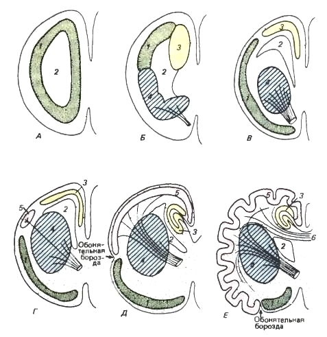
Schematic sections through the left hemisphere of the forebrain. The color codes are the same as in the previous figure.
1 - paleopallium; 2 - lateral ventricle; 3 - archipallium; 4 - basal nuclei; 5 - neopallium; 6 - corpus callosum.
A is the primitive stage. The hemisphere, in fact, is the olfactory lobe. Weakly differentiated gray matter is located inside the brain.
B - stage observed in modern amphibians. The gray matter is still located far from the outer surface, but is already subdivided into paleopallium (= olfactory lobe), archipallium (= hippocampus) and basal nuclei (= striatum). The latter takes on the significance of an associative center that has afferent and efferent connections with the thalamus (shown as lines symbolizing cut fiber bundles).
B - a more progressive stage, in which the basal nuclei sink into the hemisphere, while the sections of the cortex have moved somewhat outward.
G - the stage at which advanced reptiles are located. A neopallium appears.
D - the stage of a primitive mammal. The neopallium has increased. It has extensive connections to the brain stem. The archipallium on the medial surface of the hemisphere wraps up like a hippocampus. The paleopallium is still highly developed.
E - the stage of a highly organized mammal. The neopallium grows extremely large and gathers into folds. The paleopallium occupies a limited ventral region, representing a pear-shaped lobe. The corpus callosum develops - a powerful bridge connecting the neopallium regions of the two hemispheres. (According to Romer and Parsons, 1992.) The neopallium bark is called the new bark, or neocortex (neocortex). In mammals, it serves as the center of the higher (conditioned reflex) nervous activity coordinating the work of other parts of the brain. From here, impulses are sent to various organs and tissues of the body, and physiological processes are regulated here in accordance with environmental conditions. It is the new cortex that accumulates traces of single excitations and their combinations, as a result of which the working memory is enriched, which provides the possibility of choosing optimal solutions in new situations. Often these decisions are new combinations of previously known behavioral elements, but there is also the development and consolidation of new options for action. As it develops, the new cortex not only assumes the functions of a correlative and associative center for newly emerging types of higher nervous activity, but also begins to perform many functions that previously belonged to the centers of the brain stem and basal nuclei. At the same time, the ancient centers that control instinctive acts are not liquidated, but only subject to higher control.
In connection with the development of the neocortex, the roof of the midbrain loses its former significance, remaining only a reflex and transmission center. Auditory and other somatic sensory impulses are transmitted forward to the thalamus, most of the visual fibers are interrupted here, and all these signals from the thalamus are transmitted to the hemispheres along powerful nerve bundles. Similar thalamic connections with basal nuclei originated in low-organized groups of vertebrates and were most developed in birds. Unlike birds, in mammals the bulk of the fibers pass through the striatum through and through and diverge to the surface of the new cortex. Thus, a complete set of sensory data flows to it, on the basis of which the corresponding motor "decisions" are made in the cortex.
As already mentioned, some of the signals are transmitted from the cortex to the cerebellum through the pons and provide the necessary regulatory effects. The neocortex also has connections to the striatum and even to the hypothalamus - and thus to the autonomic nervous system. However, the bulk of the motor commands are sent along the pyramidal path (tractus corticospinalis) - a special nerve bundle that goes directly, without switching, from the cerebral cortex through the midbrain to the somatic motor areas of the brain stem and spinal cord. At the same time, the fibers of the lateral part of this path cross and innervate the opposite side of the body (i.e., the left fiber innervates the right side of the body, and vice versa), while the ventral fibers remain connected to their side of the body. The pyramidal pathway is present only in mammals, which clearly demonstrates the dominant position of the new cortex in them. greatest development this structure reaches in monkeys and, especially, in humans, playing important role in upright position. In marsupials, pyramidal axons reach only the thoracic region, while in monotremes, the pyramidal pathway is completely absent. 
The ventricles of the human brain; side view from the left side. The ventricles are shown as a cast, while the brain tissues are not shown. With the growth of the forebrain hemisphere, the lateral ventricle spread back with the formation of the posterior horn in the occipital lobe, and in its lateral part - down and forward with the formation of the lateral horn in the temporal lobe. These outgrowths, pointing backwards and downwards, led to changes in the arrangement of various parts of the brain. The hippocampus, which developed in a dorsal position on the medial surface of the hemisphere, has moved back and down to a ventral position in highly developed mammals. (According to Romer and Parsons, 1992.) Since the neocortex is a thin sheet of layered cellular material under which lies the white fibrous mass of the brain, a simple increase in the volume of the hemispheres cannot produce a proportional expansion of the cortex. At the same time, in advanced forms, the area of the bark can increase significantly due to its folding. The folds formed in this way are called convolutions (gyri), and the deep gaps between them are called furrows (sulci). Both of them contain common morphological components. In the simplest case, there is one deep Sylvian groove separating the frontal lobe (lobus frontalis) from the temporal lobe (lobus temporalis). Then, above and anterior to the Sylvian sulcus, a transverse Roland's sulcus appears, separating the frontal lobe from the parietal (lobus parietalis) from above. In primates, the transverse groove separates the small posterior occipital lobe (lobus occipitalis). In addition to the main furrows, many additional ones are formed; their number is especially high in primates and toothed whales. Previously, it was believed that the furrows in some cases indicate morphological boundaries corresponding to certain areas of the cortex. However, further studies have shown that there is no fixed relationship between the distribution of folding and the structural subdivision of the cortex (except for the nasal sulcus and, to some extent, the central sulcus in primates, which will be discussed later). It is noteworthy that the folding of the crust has developed in several evolutionary trunks of mammals quite independently. In relatively primitive mammals, such as monotremes, marsupials, and some placentals (insectivores, bats, rodents, lagomorphs), the bark is more modestly developed and has a smooth surface. 
Location of the brain in the skull of a fossil and living canid. There is a noticeable increase in the size and complication of the brain, especially the hemispheres of the forebrain. Hesperocion ( Hesperocyon gregarius) (left) is an Oligocene form that lived approximately 30 million years ago. Fenech ( Vulpes zerda) (on right) - modern form similar sizes. (According to Romer and Parsons, 1992.) The gray matter of the neocortex is characterized by a complex histological structure. In placental mammals, 6 layers of cells lying one above the other and fibers intruding between them are distinguished; this greatly distinguishes the neocortex from the remaining sections of the paleopallium and archipallium cortex, where only 2 to 4 cell layers can be distinguished. According to current estimates, in mammals with especially large brains, the number of cells in the new cortex can reach billions.
The white matter, located under the gray, in addition to the fan of connections going from the cortex to the underlying parts of the brain and back, includes a huge number of intertwining transverse fibers connecting various areas of the cortex itself. The commissure formed in this way is stretched backwards (according to the course of growth of the hemispheres) and is divided into two plates fused along the posterior edge. The lower one, thinner and deviated downward by the front edge, is the arch (fornix), the commissure of the archipallium cortex (i.e., the hippocampus). The upper, thicker, horizontally located commissure belongs to the new cortex and is called the corpus callosum (corpus callosum). This formation allows you to combine the memory of both hemispheres and significantly increases the brain's ability to learn. The corpus callosum is present only in placentals due to the significant development of the new cortex; monotremes and marsupials are deprived of it. In addition, all mammals have an anterior commissure (commissura anterior), which connects the olfactory areas of the cortex. 
The layered arrangement of nerve cells in the cortex of the telencephalon of a mammal (according to Naumov and Kartashev, 1979.) A complex system of “conductors” connecting all parts of the cortex suggests that gray matter is, in principle, a single formation, all parts of which have the same opportunities for the implementation of any functions of the cerebral hemispheres. To a certain extent, this is true: experiments show that in laboratory animals it is possible to destroy a significant part of the new cortex without causing permanent disturbances in their normal activity. Evidence of injury and morbid change confirms that this is also true for human brain. At the same time, it is clear that certain areas of the cortex are normally associated with the performance of quite specific functions. We have mentioned above the areas of the paleopallium and archipallium, intended mainly for the analysis of olfactory information and preserved, respectively, in the form of a pear-shaped lobe and a hippocampus. Differentiation of individual areas also takes place in the neopallium bark. The anterior part of the hemispheres contains the motor area. The frontal lobe located here, among other things, controls the communication of animals, including acoustic; in humans, it is associated with speech, i.e., the second signaling system. The back of the hemispheres is associated with the perception of sensations. In the occipital and temporal lobes are areas that control vision and hearing, respectively. Further forward, near the motor area, there are areas that perceive tactile and proprioceptive signals. In primates, the central sulcus (sulcus centralis), which crosses the top of the hemisphere from the medial to the lateral surface, delimits (although not quite exactly) the motor area from the sensory one. Along the anterior edge of the central sulcus, specific motor areas are located in a linear order, serving each part of the body and limbs. Along the posterior edge of the central sulcus, areas of sensory perception of the corresponding parts of the body are placed in the same order.
Thus, in many mammals, almost the entire surface of the neocortex is occupied by areas more or less closely associated with certain sensory or motor functions. Although the central sulcus may be absent, in most cases placentals have a similar linear arrangement of sensory and motor areas against each other. In marsupials (and among placentals, in xenartras), the “marking” of body areas is approximately the same, but the sensory areas are not separated from the motor areas, but are interspersed with them. But, for example, in humans, these specific functional areas occupy relatively little space on the surface of the neocortex. Between them, vast areas of gray matter have arisen (one particularly large such area occupies most of the frontal lobe), which are not associated with specific sensory or motor functions. Therefore, these areas are often referred to as "blank spots", although, as damage to these areas shows, it is in them that our higher mental abilities are located, including learning opportunities, initiative, foresight, and judgment. However, there are areas that can be removed without serious consequences for intellectual activity. 
Functional centers of the cerebral cortex of the shrew ( sorex sp.) (A) and human ( Homo sapiens
) (B) (according to Naumov and Kartashev, 1979):
1 - motor center; 2 - center of skin-muscular sensitivity; 3 - visual center; 4 - auditory center; 5 - olfactory bulb; 6 - olfactory lobes; 7 - roof of the midbrain; 8 - cerebellum; 9 - frontal lobe. The evolution of the brain is greatly influenced by external environment and motor (food-producing, defensive) activity. At the same time, the development of various parts of the brain is determined mainly by the ways of finding food: in a dog ( canis lupus), which uses the sense of smell in this process, the olfactory region is more developed; at the cat ( Felis silvestris), looking for food with the help of vision - visual; at the macaque ( Macaca mulatta), which uses vision and hearing - visual and auditory.
It is usually assumed that the size of the cerebral hemispheres determine the differences in the mental abilities of different mammals. AT in a certain sense this is true, but with significant reservations. The larger brain is made up of more nerve cells. If the area of the existing surface of the cortex is in any way connected with intelligence, then it is obvious that of two variants of the brain of the same size, the one with a furrowed surface will be more developed, and the brain with a smooth surface will be less developed. The size of the animal itself also affects the volume of the brain. This happens if only because the brain must have larger areas to serve the larger sensory and motor connections. However, the increase in brain size is not entirely proportional to body mass, so that large animals tend to have relatively smaller brains without any apparent loss of intelligence. Thus, the absolute size of the brain is not an unconditional criterion of intelligence. This is definitely indicated by the fact that the whale's brain can be five times larger in volume than the human brain.
Comparison of the brains of some mammals:
1 - horse; 2 - dog; 3 - kangaroo; 4 - person; 5 - elephant. The percentage of the brain in the total body mass is called the cephalization index. In large insectivores, it is about 0.6%, in small ones - up to 1.2%, in large cetaceans - about 0.3%, and in small ones - up to 1.7%. Most primates have a cephalization index of 1-2%. In humans, it reaches 2-3%, and some small broad-nosed monkeys have a brain, the mass of which is up to 7% of body weight. At the same time, in modern reptiles and birds, the cephalization index ranges from 0.05 to 0.5%.
Below is the mass of the brain of some mammals (the mass of the animal is indicated in brackets):
virginian opossum ( Didelphis virginiana) - 7.6 g (5 kg);
koala ( Phascolarctos cinereus) - 19.2 g (8 kg);
bush elephant ( Loxodonta africana) - 6000 g (5000 kg);
common hedgehog ( Erinaceus europaeus) - 3.3 g (1 kg);
house mouse ( Mus muscle) - 0.3 g (0.02 kg);
gray rat ( Rattus norvegicus) - 2 g (0.3 kg);
common squirrel ( Sciurus vulgaris) - 7 g (0.4 kg);
European rabbit ( Oryctolagus cuniculus) - 11 g (3 kg);
domestic horse ( Equus ferus) - 530 g (500 kg);
black rhino ( Diceros bicornis) - 500 g (1200 kg);
white-tailed deer ( Odocoileus virginianus) - 500 g (200 kg);
giraffe ( Giraffa camelopardalis) - 680 g (800 kg);
domestic sheep ( Ovis orientalis) - 140 g (55 kg);
domestic bull ( Bos primigenius) - 490 g (700 kg);
bactrian camel ( camelus bactrianus) - 762 g (700 kg);
hippo ( Hippopotamus amphibius) - 580 g (3500 kg);
white-barreled dolphin ( Delphinus delphis) - 815 g (60 kg);
narwhal ( Monodon monoceros) - 2997 g (1578 kg);
sperm whale ( Physeter macrocephalus) - 8028 g (35833 kg);
blue whale ( Balaenoptera musculus) - 3636 g (50900 kg);
domestic cat ( Felis silvestris) - 25 g (3 kg);
a lion ( panthera leo) - 270 g (250 kg);
common fox ( Vulpes vulpes) - 53 g (4.5 kg);
domestic dog ( canis lupus) - 64 g (10 kg);
polar bear ( Ursus maritimus) - 500 g (700 kg);
walrus ( Odobenus rosmarus) - 1130 g (700 kg);
marmoset Geldi ( Callimico goeldii) - 7 g (0.2 kg);
white-fronted capuchin ( Cebus albifrons) - 57 g (1 kg);
rhesus monkey ( Macaca mulatta) - 88 g (6.5 kg);
baboon ( Papio cynocephalus) - 200 g (25 kg);
silver gibbon ( Hylobates moloch) - 112 g (6.5 kg);
kalimantan orangutan ( Pongo pygmaeus) - 413 g (50 kg);
western gorilla ( gorilla gorilla) - 506 g (126 kg);
common chimpanzee ( Pan troglodytes) - 430 g (55 kg);
reasonable person ( Homo sapiens) - 1400 g (72 kg).
It can be seen from the examples given that in smaller mammals the brain is almost always relatively larger, and as the size of the body of the animal increases, the relative size of the brain decreases. This is especially pronounced among related mammalian species - for example, in a cat ( Felis silvestris) and lion ( panthera leo). Dogs are very convincing in this sense. various breeds. If the body weights of the smallest and largest breeds are approximately in the ratio 1:33, then the brain masses of the same breeds are related as 1:3. 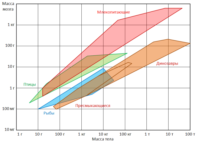
Ranges of brain and body masses for some groups of vertebrates. In domesticated animals, deprived of the need to get food and defend themselves from enemies, the size of the brain is significantly reduced. For example, the brain volume of a wolf ( canis lupus) is 30% larger than a dog of the same size. Interestingly, these changes affect not only traditionally domestic animals, but also representatives of free-living species that have been kept in captivity for some time. Yes, foxes Vulpes vulpes), born in nature, but from the very first days living in captivity, have a smaller brain than their relatives living in natural conditions. At the same time, the differences reach 20%, which approximately corresponds to the difference in brain volume between wild and real domestic animals. Brain shrinkage, although less pronounced (at about 5%), has been found in captive wolves ( Canis), ferrets ( Mustela), rats ( Rattus). At the same time, the decrease does not capture all parts of the brain, but only those areas that are associated with the work of the sense organs. The most remarkable thing is that in released pets, the weight of the brain increases. For example, feral cats have about 10% larger brains than their domestic counterparts. A significant increase in the brain was also found in feral rabbits ( Oryctolagus cuniculus) in the Kerguelen Islands. feral donkeys ( Equus asinus) in South America had 15% larger brains than domestic ones. It is also interesting that the brains of a Neanderthal ( Homo neanderthalensis) and Paleolithic Homo sapiens ( Homo sapiens) were slightly larger than the brain of a modern person.
It has been established that in many mammals motor asymmetry is observed, i.e., the predominant use of the right or left half of the body. For example, when studying unbroken horses ( Equus ferus) recorded on which foot the animals start walking, on which side they prefer to go around obstacles, and on which side they prefer to lie in a hay stall. As a result, most of the mares turned out to be right-handed, and most of the stallions were left-handed. Approximately 10% of the horses did not give preference to either the right or the left limb. According to observations, about 90% of walruses ( Odobenus rosmarus) dig mollusks out of sea silt with their right flippers. Lulling babies, about 80% of female chimpanzees ( Pan) and gorillas ( Gorilla) press their heads to the left side of their chest (approximately the same percentage is observed among women). Rats ( Rattus), leading the search for food with the help of vibrissae located on right side muzzles, are more prey than their left-handed relatives.
The cerebral hemispheres are the largest regions of the brain. In humans, the cerebral hemispheres have received maximum development compared to the rest of the parts, which largely distinguishes the human and animal brains. The left and right hemispheres of the brain are separated from each other by a longitudinal fissure running along the midline. If you look at the surface of the brain from above and from the side, you can see a slit-like depression that starts 1 cm posteriorly from the median point between the anterior and posterior poles of the brain and goes deeper. This is the central (Roland) furrow. Below it, a second large slit-lateral (Sylvian) groove passes along the lateral surface of the brain. Functions of the cerebral hemisphere of the forebrain - the topic of the article.
1 106654
Photo gallery: Functions of the cerebral hemisphere of the forebrain
Lobes of the brain
The cerebral hemispheres are divided into lobes, the names of which are given by the bones covering them:. frontal lobes located in front of the Roland and above the Sylvian furrow.
The parietal lobe lies behind the central and above the posterior portion of the lateral sulcus; it extends back to the parietal-occipital sulcus - a gap that separates the parietal lobe from the occipital one, which forms the back of the brain.
The temporal lobe is the area below the Sylvian sulcus and borders behind the occipital lobe.
Since the brain grows rapidly even before birth, the cerebral cortex begins to increase its surface, forming folds, which leads to the formation of a characteristic appearance a brain resembling a walnut. These folds are known as convolutions, and the grooves separating them are called furrows. Certain grooves in all people are located in the same place, therefore they are used as landmarks for dividing the brain into four lobes.
Development of convolutions and furrows
Furrows and convolutions begin to appear on the 3-4th month of fetal development. Up to this point, the surface of the brain remains smooth, like the brains of birds or amphibians. The formation of a folded structure provides an increase in the surface area of the cerebral cortex in a limited volume of the cranium. Different areas of the cerebral cortex perform certain, highly specialized functions. The cerebral cortex can be divided into the following areas:
Motor zones - initiate and control body movements. The primary motor area controls voluntary movements on the opposite side of the body. Directly in front of the motor area of the cortex is the so-called premotor cortex, and the third area - the additional motor area - lies on the inner surface of the frontal lobe.
Sensory areas of the cerebral cortex perceive and summarize information from sensory receptors throughout the body. The primary somatosensory area receives information from the opposite side of the body in the form of impulses from sensory receptors for touch, pain, temperature, and joint and muscle position (proprioceptive receptors).
The surface of the human body has its "representations" in the sensory and motor areas of the cerebral cortex, which are organized in a certain way. Canadian neurosurgeon Wilder Penfield, who practiced in the 1950s, created kind of map sensory zones cerebral cortex, which perceive information from various parts of the body. As part of his research, he conducted experiments in which he asked a person under local anesthesia to describe his feelings at the moment when he stimulated certain areas of the surface of the brain. Penfield found that stimulation of the postcentral gyrus produced tactile sensations in specific areas on the opposite side of the body. Other studies have shown that the volume of the motor cortex, which is responsible for various areas of the human body, depends more on the level of complexity and accuracy of the movements performed than on strength and volume. muscle mass. The cerebral cortex consists of two main layers: gray matter - a thin layer of nerve and glial cells about 2-A mm thick and white matter, which is formed by nerve fibers (axons) and glial cells.
The surface of the cerebral hemispheres is covered with a layer of gray matter, the thickness of which in different parts of the brain varies from 2 to 4 mm. Gray matter is formed by the bodies of nerve cells (neurons) and glial cells that perform a supporting function. In most of the cerebral cortex, under a microscope, six separate layers of cells can be found.
Neurons of the cerebral cortex
- Pyramidal cells got their name due to the shape of the body of the neuron, which resembles a pyramid; their axons (nerve fibers) emerge from the cerebral cortex and carry information to other parts of the brain.
- Non-pyramidal cells (all the rest) are designed to perceive and process information from other sources.
The thickness of the six layers of cells that make up the cerebral cortex varies greatly depending on the area of the brain. The German neurologist Korbinian Brodmann (1868-191) investigated these differences by staining nerve cells and examining them under a microscope. The result of Brodmann's scientific research was the division of the cerebral cortex into 50 separate sections based on certain anatomical criteria. Subsequent studies have shown that the "Brodmann fields" identified in this way play a specific physiological role and have unique ways of interacting.
Large hemispheres large hemispheres
brain, paired formations, united by the corpus callosum into the so-called telencephalon. The surface of the cerebral hemispheres is represented by numerous large or small deep convolutions. There are lobes: frontal, parietal, temporal, insular, occipital. The gray matter of the brain, consisting of nerve cells - neurons, forms the cerebral cortex and subcortical ganglia (nodes). The white matter is formed by the processes of neurons that make up the pathways of the brain.
LARGE HEMISPHERESLARGE HEMISPHERES of the brain, paired formations united by the corpus callosum (cm. corpus callosum) in so-called. terminal brain. The surface of the cerebral hemispheres is represented by numerous large or small deep convolutions. There are lobes: frontal, parietal, temporal, insular, occipital. The gray matter of the brain, consisting of nerve cells - neurons, forms the cerebral cortex and subcortical ganglia (cm. GANGLION)(nodes). The white matter is formed by the processes of neurons that make up the pathways of the brain.
encyclopedic Dictionary . 2009 .
See what the "large hemispheres" are in other dictionaries:
The brain is paired formations, united by the corpus callosum in the so-called. terminal brain. The surface of the cerebral hemispheres is represented by numerous large or small deep convolutions. There are lobes: frontal, parietal, temporal, insular... Big Encyclopedic Dictionary
The brain, paired formations, united by the corpus callosum in the so-called. terminal brain. The surface of the B. p. is represented by numerous. b. or m. deep convolutions. There are lobes: frontal, parietal, temporal, insular, occipital. Gray in ... ... Natural science. encyclopedic Dictionary
LARGE HEMISPHERES OF THE BRAIN- the higher parts of the brain, consisting of the surface layer of the cerebral cortex and the deep parts of the subcortex; cover the cerebellum and brain stem. B. p. g. m. are divided along the midline into the right and left hemisphere that are in the depths... Psychomotor: Dictionary Reference
The Arctic Ocean, in contrast to the South, represents a completely Mediterranean character. It has natural boundaries for a considerable distance and only in three places directly merges with the waters of the Atlantic and Pacific ... ...
The Arctic Ocean, in contrast to the southern one, is completely mediterranean in character. It has natural boundaries for a considerable distance and only in three places directly merges with the waters of the Atlantic and Pacific ... ... Encyclopedic Dictionary F.A. Brockhaus and I.A. Efron
Also called comparative morphology, this is the study of the patterns of structure and development of organs by comparing various kinds Living creatures. Comparative anatomy data traditional basis biological classification. Under the morphology... Collier Encyclopedia
Sectional view of the brain of an adult male. The human brain (Latin encephalon) is about ... Wikipedia
The science that studies the structure of the body, individual organs, tissues and their relationships in the body. All living things are characterized by four features: growth, metabolism, irritability and the ability to reproduce themselves. The combination of these signs ... ... Collier Encyclopedia
Animals (Mammalia), a class of vertebrates, the most famous group animals, including more than 4600 species of world fauna. It includes cats, dogs, cows, elephants, mice, whales, people, etc. In the course of evolution, mammals have carried out the widest ... ... Collier Encyclopedia
I Medicine Medicine is a system of scientific knowledge and practice aimed at strengthening and maintaining health, prolonging people's lives, and preventing and treating human diseases. To accomplish these tasks, M. studies the structure and ... ... Medical Encyclopedia