Anatomy and physiology of the heart: structure, functions, hemodynamics, cardiac cycle, morphology
The structure of the heart of any organism has many characteristic nuances. In the process of phylogenesis, that is, the evolution of living organisms to more complex ones, the heart of birds, animals and humans acquires four chambers instead of two chambers in fish and three chambers in amphibians. Such a complex structure the best way adapted to separate arterial and venous blood flows. In addition, the anatomy of the human heart involves many the smallest details, each of which performs its strictly defined functions.
Heart as an organ
So, the heart is nothing more than a hollow organ consisting of specific muscle tissue, which carries out the motor function. The heart is located in chest behind the sternum, more to the left, and its longitudinal axis is directed anteriorly, to the left and down. In front, the heart borders on the lungs, almost completely covering them, leaving only a small part directly adjacent to the chest from the inside. The boundaries of this part are otherwise called absolute cardiac dullness, and they can be determined by tapping the chest wall ().
In people with a normal constitution, the heart has a semi-horizontal position in the chest cavity, in people with an asthenic constitution (thin and tall) it is almost vertical, and in hypersthenics (dense, stocky, with a large muscle mass) – almost horizontal.

heart position
The posterior wall of the heart is adjacent to the esophagus and to the large main vessels (thoracic aorta, inferior vena cava). The lower part of the heart is located on the diaphragm.

external structure of the heart
Age characteristics
The human heart begins to form in the third week of the intrauterine period and continues throughout the entire period of gestation, passing through stages from a single-chamber cavity to a four-chamber heart.

development of the heart in utero
The formation of four chambers (two atria and two ventricles) occurs already in the first two months of pregnancy. The smallest structures are fully formed by birth. It is in the first two months that the heart of the embryo is most vulnerable to the negative influence of certain factors on the expectant mother.
The fetal heart participates in the blood flow throughout its body, but differs in the circles of blood circulation - the fetus does not yet have its own breathing with lungs, and it “breathes” through placental blood. There are some openings in the fetal heart that allow pulmonary blood flow to be “switched off” from the circulation before birth. During childbirth, accompanied by the first cry of the newborn, and, consequently, at the moment of increased intrathoracic pressure and pressure in the baby's heart, these openings close. But this does not always happen, and the child may still have them, for example (not to be confused with a defect such as atrial septal defect). An open window is not a heart defect, and subsequently, as the child grows, it closes.

hemodynamics in the heart before and after birth
The heart of a newborn baby has a round shape, and its dimensions are 3-4 cm in length and 3-3.5 cm in width. In the first year of a child's life, the heart increases significantly in size, more in length than in width. The weight of a newborn baby's heart is about 25-30 grams.
As the baby grows and develops, the heart also grows, sometimes significantly ahead of the development of the body itself according to age. By the age of 15, the mass of the heart increases almost tenfold, and its volume increases more than fivefold. The heart grows most rapidly until the age of five, and then during puberty.
In an adult, the size of the heart is about 11-14 cm in length and 8-10 cm in width. Many people rightly believe that the size of each person’s heart corresponds to the size of his clenched fist. The weight of the heart in women is about 200 grams, and in men it is about 300-350 grams.
After age 25, changes begin in the connective tissue of the heart, which forms the heart valves. Their elasticity is no longer the same as in childhood and adolescence, and the edges may become uneven. As a person grows and then ages, changes occur in all structures of the heart, as well as in the vessels that feed it (the coronary arteries). These changes can lead to the development of numerous cardiac diseases.
Anatomical and functional features of the heart
Anatomically, the heart is an organ divided into four chambers by septa and valves. The “upper” two are called atria (atrium), and the “lower” two are called ventricles (ventriculum). Between the right and left atria is the interatrial septum, and between the ventricles is the interventricular septum. Normally, these septa do not have holes in them. If there are holes, this leads to mixing of arterial and venous blood, and, accordingly, to hypoxia of many organs and tissues. Such holes are called septal defects and are classified as.

basic structure of the chambers of the heart
The boundaries between the upper and lower chambers are the atrioventricular openings - the left one, covered by the mitral valve leaflets, and the right one, covered by the tricuspid valve leaflets. The integrity of the septa and the proper operation of the valve leaflets prevent the mixing of blood flows in the heart and promote clear unidirectional blood flow.
The atria and ventricles are different - the atria are smaller than the ventricles and have thinner walls. Thus, the wall of the atria is about only three millimeters, the wall of the right ventricle is about 0.5 cm, and the wall of the left is about 1.5 cm.
The atria have small projections called ears. They have a slight suction function for better pumping of blood into the atrium cavity. The mouth of the vena cava flows into the right atrium near its appendage, and four (less often five) pulmonary veins flow into the left atrium. The pulmonary artery (more often called the pulmonary trunk) on the right and the aortic bulb on the left depart from the ventricles.

structure of the heart and its vessels
From the inside, the upper and lower chambers of the heart are also different and have their own characteristics. The surface of the atria is smoother than the ventricles. Thin connective tissue valves originate from the valve ring between the atrium and the ventricle - bicuspid (mitral) on the left and tricuspid (tricuspid) on the right. The other edge of the valves faces the inside of the ventricles. But so that they do not hang freely, they are supported, as it were, by thin tendon threads called chords. They are like springs, stretch when the valve flaps close and compress when the valve flaps open. The chordae originate from the papillary muscles from the wall of the ventricles - three in the right and two in the left ventricle. That is why the ventricular cavity has an uneven and lumpy inner surface.
The functions of the atria and ventricles also differ. Due to the fact that the atria need to push blood into the ventricles, and not into larger and longer vessels, they have to overcome less resistance from muscle tissue, therefore the atria are smaller in size and their walls are thinner than those of the ventricles. The ventricles push blood into the aorta (left) and the pulmonary artery (right). Conventionally, the heart is divided into right and left halves. The right half serves for the flow of exclusively venous blood, and the left half for arterial blood. Schematically, the “right heart” is indicated in blue, and the “left heart” is indicated in red. Normally, these flows never mix.

hemodynamics in the heart
One cardiac cycle lasts about 1 second and is carried out as follows. At the moment the atria are filled with blood, their walls relax - atrial diastole occurs. The valves of the vena cava and pulmonary veins are open. The tricuspid and mitral valves are closed. Then the atrial walls tense and push blood into the ventricles, the tricuspid and mitral valves are open. At this moment, systole (contraction) of the atria and diastole (relaxation) of the ventricles occur. After the ventricles receive blood, the tricuspid and mitral valves close, and the aortic and pulmonary valves open. Next, the ventricles contract (ventricular systole), and the atria fill with blood again. The general diastole of the heart begins.

cardiac cycle
The main function of the heart is reduced to pumping, that is, to pushing a certain blood volume into the aorta with such pressure and speed that the blood is delivered to the most distant organs and to the smallest cells of the body. Moreover, arterial blood with a high content of oxygen and nutrients is pushed into the aorta, entering the left half of the heart from the vessels of the lungs (flows to the heart through the pulmonary veins).
Venous blood, with low content oxygen and other substances are collected from all cells and organs from the vena cava system, and flow into the right half of the heart from the superior and inferior vena cava. Next, venous blood is pushed from the right ventricle into the pulmonary artery, and then into the pulmonary vessels in order to carry out gas exchange in the alveoli of the lungs and to enrich it with oxygen. In the lungs, arterial blood collects in the pulmonary venules and veins, and again flows into the left side of the heart (the left atrium). And so the heart regularly pumps blood throughout the body at a frequency of 60-80 beats per minute. These processes are designated by the concept "Circles of Blood Circulation". There are two of them - small and large:
- Small circle includes the flow of venous blood from the right atrium through the tricuspid valve into the right ventricle - then into the pulmonary artery - then into the arteries of the lungs - oxygenation of blood in the pulmonary alveoli - flow of arterial blood into the smallest veins of the lungs - into the pulmonary veins - into the left atrium.
- Big circle includes the flow of arterial blood from the left atrium through the mitral valve into the left ventricle - through the aorta into the arterial bed of all organs - after gas exchange in tissues and organs, the blood becomes venous (with a high content carbon dioxide instead of oxygen) - further into the venous bed of the organs - into the system of vena cava - into the right atrium.

circulation circles
Video: cardiac anatomy and cardiac cycle briefly
Morphological features of the heart
If you examine sections of the heart under a microscope, you can see a special type of muscle that is not found in any other organ. This is a type of striated muscle, but has significant histological differences from ordinary skeletal muscles and from the muscles lining the internal organs. The main function of the heart muscle, or myocardium, is to provide the most important ability of the heart, which forms the basis for the vital activity of the entire organism as a whole. This is the ability to contract, or contractility.In order for the heart muscle fibers to contract synchronously, electrical signals must be supplied to them, which excite the fibers. This is another ability of the heart – .
Conduction and contractility are possible due to the fact that the heart autonomously generates electricity. Function data (automatism and excitability) are provided by special fibers that are integral part conducting system. The latter is represented by electrically active cells of the sinus node, atrioventricular node, the bundle of His (with two legs - right and left), as well as Purkinje fibers. In the case when a patient’s myocardial damage affects these fibers, they develop, otherwise called.
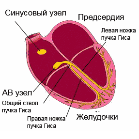
cardiac cycle
Normally, the electrical impulse originates in the cells of the sinus node, which is located in the area of the right atrium appendage. In a short period of time (about half a millisecond), the impulse spreads throughout the atrial myocardium and then enters the cells of the atrioventricular junction. Typically, signals are transmitted to the AV node through three main tracts - the Wenkenbach, Thorel and Bachmann bundles. In the cells of the AV node, the impulse transmission time is extended to 20-80 milliseconds, and then the impulses travel through the right and left branches (as well as the anterior and posterior branches of the left branch) of the His bundle to the Purkinje fibers, and ultimately to the working myocardium. The frequency of impulse transmission along all pathways is equal to the heart rate and is 55-80 impulses per minute.
So, the myocardium, or cardiac muscle, is the middle layer in the wall of the heart. The inner and outer membranes are connective tissue and are called the endocardium and epicardium. The last layer is part of the pericardial sac, or cardiac “shirt”. Between the inner layer of the pericardium and the epicardium, a cavity is formed, filled with a very small amount of fluid, to ensure better sliding of the pericardial layers during heart contractions. Normally, the fluid volume is up to 50 ml; exceeding this volume may indicate pericarditis.

structure of the heart wall and membrane
Blood supply and innervation of the heart
Despite the fact that the heart is a pump to supply the entire body with oxygen and nutrients, it itself also needs arterial blood. In this regard, the entire wall of the heart has a well-developed arterial network, which is represented by the branching of the coronary (coronary) arteries. The orifices of the right and left coronary arteries depart from the root of the aorta and are divided into branches that penetrate into the thickness of the heart wall. If these important arteries become clogged with blood clots and atherosclerotic plaques, the patient will develop and the organ will no longer be able to perform its functions fully.

location of the coronary arteries supplying blood to the heart muscle (myocardium)
The frequency and force with which the heart beats is influenced by nerve fibers extending from the most important nerve conductors - the vagus nerve and the sympathetic trunk. The first fibers have the ability to slow down the rhythm frequency, the latter - to increase the frequency and strength of the heartbeat, that is, they act like adrenaline.
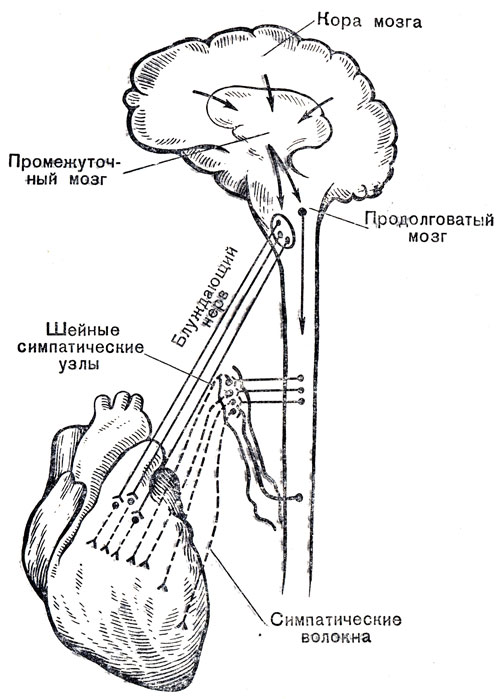
innervation of the heart
In conclusion, it should be noted that the anatomy of the heart may have any deviations in individual patients, therefore, only a doctor can determine the norm or pathology in a person after conducting an examination that can most informatively visualize the cardiovascular system.
Video: lecture on cardiac anatomy
When there is eternal spring in the soul, there is no way to contain the high spirits: it is bursting from the chest to splash out with sweet creativity. How to draw, or better yet, both together? Take a simple pencil Blank sheet paper - now you will find out everything.
Lesson #1: How to draw a heart with a pencil
We will draw a heart of roses. Draw a regular circle and divide it in half with a line. Right on horizontal line draw an uneven oval, similar to a deflated balloon. Attach a couple of curves to it at the top and bottom, like the red lines shown in the example figure.

In the lesson explaining how to draw a heart, first of all, pay attention to the red lines of the sample - these are new fragments that need to be repeated on your version of the original heart.

Draw some kind of snail in the very center of the future masterpiece. First, simply divide the uneven oval almost in half with a convex curved line. Add a few strokes: in the form of the letter “P” and from its top regular line, bounded by the same oval. Don't forget to add one more stroke, very small, in the upper petal. This inverted “comma” will add volume to the drawing.

A very easy step creative process entitled “How to draw a heart”: draw two symmetrical petals, the upper part of which follows the invisible lines of the heart.

All that's left is to add three petals at the bottom of the heart. If you didn't skip math classes at school, then you know how to draw curly braces. This acquired skill will help you easily cope with the task: a couple of curves on the left and right and another, final one, with an “arrow” down the center. By the way, if you look closely at the sample, you will see that the last petal you drew will be boring without a small detail - a convex stroke that adds volume.

Erase all auxiliary, erroneous and unnecessary lines. Lesson “How to draw a heart” is completed!
Lesson #2: Heart surrounded by roses
Let’s complicate the task: let’s draw in a scarlet round dance:

Draw a random outline of the heart, for example, like this:

Make the first sketches of three buds at once, distributing them evenly. Start with curls, draw side lines from them according to the pattern:

Each flower has its own individual uniform, which appears thanks to simple curved lines. Take a closer look and repeat them in your drawing, there is nothing complicated about it:

Let's complete the drawing of the roses by adding three or four graceful outlines to each bud.
In order to ensure adequate nutrition of internal organs, the heart pumps an average of seven tons of blood per day. Its size is equal to clenched fist. Throughout life, this organ makes approximately 2.55 billion beats. The final formation of the heart occurs by the 10th week of intrauterine development. After birth, the type of hemodynamics changes dramatically - from feeding on the mother’s placenta to independent, pulmonary breathing.
Read in this article
Muscle fibers (myocardium) are the predominant type of heart cells. They make up its bulk and are located in the middle layer.
The outside of the organ is covered with epicardium. It wraps at the level of the attachment of the aorta and pulmonary artery, heading downwards. In this way, the pericardial sac is formed. It contains about 20 - 40 ml of clear liquid, which prevents the leaves from sticking together and being injured during contractions.
The inner membrane (endocardium) folds in half at the transition of the atria into the ventricles, the mouths of the aortic and pulmonary trunk, forming valves.
- Their valves are attached to a ring of connective tissue, and the free part moves with the blood flow. In order to prevent the parts from everting into the atrium, threads (chords) are attached to them, extending from the papillary muscles of the ventricles.
- The heart has the following structure:
- three membranes - endocardium, myocardium, epicardium;
- pericardial sac;
- chambers with arterial blood - left atrium (LA) and ventricle (LV);
- sections with venous blood - right atrium (RA) and ventricle (RV);
- valves between the LA and LV (mitral) and tricuspid on the right;
- two valves separate the ventricles and large vessels (aortic on the left and pulmonary artery on the right);
- the septum divides the heart into right and left halves;
efferent vessels, arteries - pulmonary (venous blood from the pancreas), aorta (arterial from the left ventricle);
afferent veins - pulmonary (with arterial blood) enter the LA, vena cava flow into the RA. In general, the LV is more powerful (compared to the right), as it forces blood into the arteries, overcoming the high resistance of the vascular walls. The PP is more developed than the left, it receives blood from the whole body, and the left one only from the lungs.
Which side of a person's heart is on?
In humans, the heart is located on the left side in the center of the chest. The main part is located in this area - 75% of the total volume. One third extends beyond the midline into the right half. In this case, the axis of the heart is inclined (oblique direction). This situation is considered classic, as it occurs in the vast majority of adults. But options are also possible:
- dextrocardia (right side);
- almost horizontal - with a wide, short chest;
- close to vertical - for thin people.
Where is a person's heart located?
The human heart is located in the chest between the lungs. It is adjacent to the sternum from the inside, and is limited below by the diaphragm. It is surrounded by the pericardium, the pericardium. Pain in the heart area appears on the left near the mammary gland. The top is projected there. But with angina, patients feel pain behind the sternum, and it spreads along the left side of the chest.
Where is the heart located in the human body?
The heart in the human body is located in the center of the chest, but its main part goes into the left half, and only one third is located on the right side. For most it has an angle of inclination, but for fat people its position is closer to horizontal, and for thin people it is closer to vertical.
Location of the heart in the human chest
In humans, the heart is located in the chest in such a way that its anterior and lateral surfaces are in contact with the lungs, and its posterior and inferior surfaces are in contact with the diaphragm. The base of the heart (from above) passes into large vessels - the aorta, pulmonary artery. The apex is the lowest part, it approximately corresponds to the 4-5 space between the ribs. It can be found in this area by lowering an imaginary perpendicular from the center of the left collarbone.
The external structure of the heart refers to its chambers; it contains two atria and two ventricles. They are separated by partitions. The pulmonary veins, the vena cava, enter the heart, and the arteries of the lungs, the aorta, carry the blood out. Between the large vessels, at the border of the atria and ventricles of the same name, there are valves:
- aortic;
- pulmonary artery;
- mitral (left);
- tricuspid (between the right parts).
The heart is surrounded by a cavity containing a small amount of fluid. It is formed by the pericardial layers.
If you clench your fist, you can imagine exactly the appearance of a heart. In this case, the part that is located at the wrist joint will be its base, and the acute angle between the first and thumb will be its apex. What is important is that its size is also very close to a clenched fist.
 This is what a human heart looks like
 Borders of the heart and their projection onto the surface of the chest
The boundaries of the heart are found by percussion, by tapping; radiography or echocardiography helps to determine them more accurately. The projections of the cardiac contour onto the surface of the chest are:
- right – 10 mm to the right of the sternum;
- left – 2 cm inward from the perpendicular from the center of the collarbone;
- apex – 5th intercostal space;
- base (upper) – 3rd rib.
What tissues make up the heart?
The composition of the heart includes the following types fabrics:
- muscle - the main one, is called the myocardium, and the cells are cardiomyocytes;
- connective – valves, chords (threads that hold the valves), outer (epicardial) layer;
- epithelium – inner lining (endocardium).

Surfaces of the human heart
The human heart has the following surfaces:
- ribs, sternum – anterior;
- pulmonary – lateral;
- diaphragmatic – lower.
Apex and base of the heart
The apex of the heart is directed down and to the left, its localization is the 5th intercostal space. It represents the top of the cone. The wide part (base) is located on top, closer to the collarbones, and is projected at the level of the 3rd rib.
Human heart shape
Heart shaped healthy person looks like a cone. Its tip is directed under acute angle down and to the left of the center of the sternum. The base contains the mouths of large vessels and is located at the level of the 3rd rib.
Right atrium
Receives blood from the vena cava. Next to them is the foramen ovale, which connects the RA and LA in the fetal heart. In a newborn, it closes after the pulmonary blood flow opens, and then completely heals. During systole (contraction), venous blood passes into the pancreas through the tricuspid valve. The RA has a fairly powerful myocardium and a cubic shape.
Left atrium
Arterial blood from the lungs passes into the LA through 4 pulmonary veins and then flows through the opening into the LV. The walls of the LA are 2 times thinner than those of the right one. The shape of the LP is similar to a cylinder.
Right ventricle
It looks like an inverted pyramid. The capacity of the pancreas is about 210 ml. It can be divided into two parts - the arterial (pulmonary) cone and the ventricular cavity itself. In the upper part there are two valves: the tricuspid and the pulmonary trunk.
Left ventricle
It looks like an inverted cone, its lower part forms the top of the heart. The thickness of the myocardium is the largest - 12 mm. There are two openings at the top - for connection with the aorta and LA. Both of them are covered by valves - the aortic and mitral.
Why are the walls of the atria thinner than the walls of the ventricles?
The thickness of the walls of the atrium is less, they are thinner, since they only need to push blood into the ventricles. The right ventricle follows them in strength; it throws its contents into the neighboring lungs, and the left one is the largest in terms of the size of its walls. It pumps blood to the aorta, where there is high pressure.
Tricuspid valve
The right atrioventricular valve consists of a sealed ring that limits the opening and leaflets; there may be not 3, but from 2 to 6.
Half of the people have a tricuspid configuration.
The function of this valve is to prevent the reflux of blood into the RA during RV systole.
Pulmonary valve
It prevents blood from passing back into the pancreas after it contracts. The composition contains valves similar in shape to a crescent. In the middle of each there is a knot that seals the closure.
Mitral valve
It has two doors, one is in the front and the other is in the back. When the valve is open, blood flows from the LA to the LV. When the ventricle contracts, its parts close together to allow blood to pass into the aorta.
Aortic valve
Formed by three semilunar-shaped flaps. Like the pulmonary one, it does not contain threads that hold the valves in place. In the area where the valve is located, the aorta expands and has depressions called sinuses.
Adult heart weight
Depending on the physique and total body weight, the weight of the heart in an adult varies from 200 to 330 g. In men, it is on average 30-50 g heavier than in women.
Diagram of blood circulation
Gas exchange occurs in the alveoli of the lungs. They receive venous blood from the pulmonary artery emerging from the pancreas.
Despite the name, the pulmonary arteries carry venous blood. After the release of carbon dioxide and oxygen saturation through the pulmonary veins, the blood passes into the left atrium. This is how a small circle of blood flow, called pulmonary, is formed.
The large circle covers the entire body as a whole. From the LV, arterial blood spreads to all vessels, nourishing the tissues. Deprived of oxygen, venous blood flows from the vena cava into the RA, then into the RV. The circles close together, ensuring a continuous flow. They are named so because of the shape of the branches, reminiscent of a crown (crown). Venous blood from the heart muscle predominantly enters the coronary sinus. It opens into the right atrium. This circle of blood circulation is considered the third, coronary.
Watch the video about the structure of the human heart:
What is special about the structure of a child’s heart?
Until the age of six, the heart is spherical due to the large atria. Its walls are easily stretched, they are much thinner than those of adults. A network of tendon threads is gradually formed, fixing the valve leaflets and papillary muscles. Full development of all heart structures ends by age 20.
The position of the newborn's heart in the chest is initially oblique, adjacent to the anterior surface. This is caused by an increase in the volume of lung tissue and a decrease in the mass of the thymus gland.
Until two years of age, the heart impulse forms the right ventricle, and then part of the left. The atria are the leaders in growth rate up to 2 years, and the ventricles after 10 years. Up to ten years, the LV is ahead of the right.
Basic functions of the myocardium
The heart muscle differs in structure from all others, as it has several unique properties:
- Automatism is excitation under the influence of one’s own bioelectric impulses. They first form in the sinus node. He is the main pacemaker, generating about 60 - 80 signals per minute. The underlying cells of the conducting system are nodes of the 2nd and 3rd order.
- Conduction - impulses from the site of formation can spread from the sinus node to the RA, LA, atrioventricular node, and along the ventricular myocardium.
- Excitability - in response to external and internal stimuli, the myocardium is activated.
- Contractility is the ability to contract when excited. This function creates the pumping capabilities of the heart. The force with which the myocardium reacts to an electrical stimulus depends on the pressure in the aorta, the degree of stretching of the fibers in diastole, and the volume of blood in the chambers.
The functioning of the heart goes through three stages:
- Contraction of the RA, LA and relaxation of the RV and LV with the opening of the valves between them. Transition of blood into the ventricles.
- Ventricular systole - the valves of the blood vessels open, blood flows into the aorta and pulmonary artery.
- General relaxation (diastole) - blood fills the atria and presses on the valves (mitral and tricuspid) until they open.
During the period of contraction of the ventricles, the valves between them and the atria are closed by blood pressure. In diastole, the pressure in the ventricles drops, it becomes lower than in large vessels, then parts of the pulmonary and aortic valves close so that the blood flow does not return.
Heart cycle
There are 2 stages in the heart cycle: contraction and relaxation. The first is called systole and also includes 2 phases:
- compression of the atria to fill the ventricles (lasts 0.1 sec.);
- the work of the ventricular part and the release of blood into large vessels (about 0.5 sec.).
Then comes relaxation - diastole (0.36 sec). Cells change polarity to respond to the next impulse (repolarization), and the blood vessels of the myocardium bring nutrition. During this period, the atria begin to fill.
The heart ensures the movement of blood through the large and small circles thanks to the coordinated work of the atria, ventricles, great vessels and valves. The myocardium has the ability to generate an electrical impulse and conduct it from the nodes of automaticity to the cells of the ventricles. In response to a signal muscle fibers
become active and contract. The cardiac cycle consists of a systolic and a diastolic period.
Useful video
Watch the video about the work of the human heart:
Read also


If any abnormality is suspected, a heart x-ray is prescribed. It can reveal a normal shadow, an increase in the size of an organ, and defects. Sometimes radiography with contrast of the esophagus is performed, as well as in one to three and sometimes even four projections.
Step 1. Okay let's start this lesson on the human heart shall we? First draw out some guidelines and shapes so we have a nice workable wireframe to use. Start with a circle for the heart and then draw the bottom of the heart which contains the heart muscle. The three horizontal lines you see drawn will be the plugs on the pulmonary artery, pulmonary veins and left atrium. The lump you see drawn will be for the aorta.

Step 2. Okay, let's draw the actual shape of the aorta, as well as the pipes that come from this part of the heart. You will then draw out the shape for the pulmonary artery like you see here.

Step 3. Now, sketch out the shape of the vena cava that is in the vase while looking at the tube, which is all self-explanatory. The next draw is four pipes. The top tube enters the pulmonary artery, and the last three are the pulmonary veins, which are on the left side. Next sketch out the outer shape of the heart on the left side, which is also part of the heart muscle, and then draw in the veins that lie on the surface of the heart. Finally you will draw the tube for the inferior vena cava, which is located just below the lower left side of the heart.

Step 4. It's yours last step drawing and all you have to do is draw out the remaining actual shape human heart, and then draw into the superficial veins. Lastly, draw the tubes for the pulmonary vein and left atrium. Erase all the guidelines that are visible to clear your drawing of the human heart.
Many of us took up a pen or pencil not only to pass homework in drawing at school. Sometimes, for one reason or another, an inexplicable urge to draw appears in the life of a teenager or an adult. How I want to pick up a pencil and simply start creating little masterpieces, even if only for myself or for a close circle of people without pretensions to global recognition and glory. It may seem that those performing simple pencil movements in the video or in front of you are putting in little to no effort, but in reality this is not the case. Professionalism in drawing, as in any other craft, comes only with experience. Even in the simplest drawings you can highlight subtleties and details that you had never even thought about before. Now we will look at one of the most simple drawings- heart. Remember school years or those moments when we all drew it to each other. This time we will learn how to draw a regular heart, with an arrow or wings. Also recommends subscribing. This way you will see new materials first.
Simple ways
And so, let's figure out how to beautifully draw a heart with a pencil step by step for beginners. All we need is a piece of paper, a simple pencil, and, of course, due perseverance in this endeavor. To make the heart cute, you need to make it symmetrical, and to do this, do a couple of simple steps:
Draw two identical circles on a piece of paper in the same horizontal plane so that both circles intersect slightly. Let’s immediately say that the upper halves of the circles will help create beautiful symmetrical edges of the heart. Accordingly, those parts that make up the main drawing can be drawn boldly, and those that need to be removed can be made weaker. It is advisable to draw circles by hand, it’s okay if at first the circle doesn’t turn out very round, this will improve with practice. But if you are not initially satisfied with the quality of the circles, you can resort to auxiliary means.
The next figure in the figure is a cross. The vertical line of the cross should pass along the intersection of the circles; to form it, it is enough to draw a line through two points at which the circles from the first point intersected. It doesn’t make sense to raise the vertical line high in length; its lower part is more useful for the drawing, so don’t skimp and lower it down. In order to understand how low you should go, ask yourself: how to draw a heart beautifully, what proportions in height will be optimal for you so that the drawing turns out beautiful. The horizontal line is drawn perpendicular to the vertical in the middle of both circles. 
From the extreme points of intersection of the circles with the horizontal line, lower two smooth symmetrical lines to the bottom point of the heart. You should determine the position of this lowest point yourself, because because of this
parameter the heart will turn out to be more elongated or more flattened. 
Draw a thick line of semicircles of each circle upward from the horizontal line and to the first point of intersection. 
At this stage, drawing the heart is complete. All that remains is to remove the extra lines used in the construction and create the resulting drawing.

The perfect heart in terms of symmetry and shape is already in front of you. Of course, this is not the only way to draw a heart.
An easier option for advanced artists
If the previous version seemed boring and unattractive to you due to the presence large quantity additional constructions, if you need to complete the drawing much faster and there is no way to build circles, if you feel you have a sufficient level and skills, we bring to your attention the second method of how to draw a heart with a pencil step by step. But let’s make a reservation right away: you must be good at drawing symmetrical circles, otherwise the heart will turn out to be asymmetrical.
- Divide the sheet into four parts with two perpendicular lines, in other words, draw the same cross.
- Mark on vertical line the position of the upper and lower points of the heart, and on the horizontal the same segment to the left and right of the intersection point.
- Connect the top point with a smooth semicircular line to the leftmost point on the horizontal axis and the same smooth semicircular line with the right point.
- From the extreme left and right points, lower two smooth symmetrical lines to the bottom edge.
For more experienced artists
The next method of depicting a heart is even simpler; it will help to depict a heart in just a couple of steps and with the effect of rotation around its axis. But this method is only suitable for experienced specialists who can easily draw symmetrical semicircular lines by hand, without using additional constructions.
- draw the most ordinary oval, the edges of which are elongated in a horizontal plane.
- divide the oval with a line in the middle; if the heart should turn out at an angle, the line should be curved in the desired direction. This frame will show you how to draw a heart step by step and quickly.
- choose a point just below the top point of the oval on a vertical line and, starting from this position, draw two lines on the top of the heart. These lines can completely fit into the oval, or they can protrude beyond its limits, it all depends on your wishes and vision perfect shape for drawing.
- repeat the previous point with the bottom of the heart - lower two symmetrical lines to the bottom point.
- add Cupid's arrow.
As a result, we get the following picture: 
Let's add details
The drawing can be supplied with additional effects, such as wings, horns, halos, inscriptions, fire and similar additions that give additional effects and allowing you to harmoniously include the image in one or another motif of the drawing, depending on your ideas. Today we will look at several options for drawing hearts with wings, as the most romantic version of this image. Wings give the hearts a special romanticism and sublime tones. It should be noted that the position of the wings in relation to the heart determines the nature of what the author wants to convey: raised to the top, spread wings show strong intentions, pure feelings, desire for a loved one. On the contrary, the more the wings go down (and possibly connect downwards), the more this shows the heart’s attempt to close itself off from some external factors and problems, an attempt to hide something under its care and care.
Wings on the heart will tell a lot
So, let's look at how to draw a heart with wings step by step with a pencil. Such a drawing will require you to first study the topic of how to draw a heart or a ready-made template with an image of a heart. So, to begin with, we start from the fact that we already have a ready-made drawing. It is clear that the simplest and most uncomplicated way is to draw the wings by hand without any frames or additional constructions. This method may seem to be the most common of the above, but at the same time the most difficult, since it will require the author to have practical skills in drawing symmetrical lines and curvilinear figures by hand. Several options for depicting wings from the heart should be highlighted. Wings can be depicted from the sides or from the top. The position of the wings themselves in this case does not matter at all; what matters is from which part they grow, so to speak.
Wings with feathers in one row
If you decide to depict wings growing from the top of the heart, then it is better to depict them as small, decorative, this gives a certain sophistication and sophistication when in visual contact with the picture. When depicting wings on the sides of the heart, excellent option there will be wings spread to the sides. We do the following:


Chic wings with feathers in several rows
If you want to get a heart effect with huge wings, a span reminiscent of a flying eagle, then it is better to use a multi-level version of the wings rather than a single-level one. The more rows of feathers there are on the wings, the more spectacular the design will seem and the nobler the impulse of the heart itself, as if an eagle’s wings are carrying it towards its lover. So, let's figure out step by step how to draw a heart with wings with a pencil, depicting a rich pattern of feathers or other additional effects on the wings.
As in previous cases, we begin the drawing with the most ordinary frame of the future heart. He can be portrayed as different ways, but it is better to choose one of the above.
To the frame of the heart itself or its finished drawing we add the frame of future wings. There is no need to skimp on space here; the span and size of the wings themselves should be truly royal. Don’t skimp on space; it’s better to draw a smaller heart. Form the frame with several levels at once: the smallest is closest to the heart, the largest is the farthest. 


Then start drawing each feather, starting with the smallest at the base and ending with the longest and largest at the edges. In principle, you can start applying layers, gradually depicting each row of feathers separately, superimposing the next one on top of it without a frame. Repeat the steps described with the second wing.